Magnetic resonance imaging
From MEpedia, a crowd-sourced encyclopedia of ME and CFS science and history
(Redirected from MRI)
Magnetic Resonance Imaging (MRI) uses magnetic fields and radio waves to produce images of thin slices of tissues. MRI scans can be used to image many different parts of the body, including the brain, joints, major organs and even the whole body.[1]
MRI scans can be used for many different purposes, e.g. to show:
- abnormalities of the brain and spinal cord
- abnormalities in various parts of the body such as breast, prostate, and liver
- joint injuries or abnormalities, for example a knee injury
- heart structure and function
- areas of activity within the brain, using a functional MRI
- blood flow through blood vessels and arteries[2]
ME/CFS MRI evidence[edit | edit source]
- Possible white matter abnormalities of unknown etiology are found on MRIs of some ME/CFS patients. These are identified by T2 hyperintensities, which might indicate lesions or Virchow-Robin spaces.[3][4]

Cost and availability[edit | edit source]
Notable studies[edit | edit source]
- 1993, A controlled study of brain magnetic resonance imaging in patients with the chronic fatigue syndrome[3]
- 1999, Brain MRI abnormalities exist in a subset of patients with chronic fatigue syndrome[4]
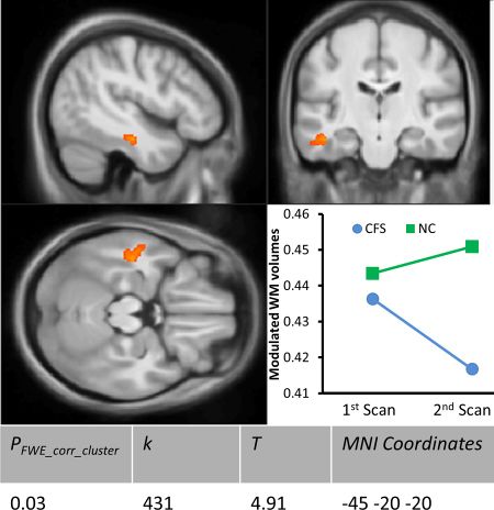
- 2016, Progressive Brain Changes in Patients With Chronic Fatigue Syndrome: A Longitudinal MRI Study[5]
- 2016, Autonomic correlations with MRI are abnormal in the brainstem vasomotor centre in Chronic Fatigue Syndrome[6]
- 2017, Medial prefrontal cortex deficits correlate with unrefreshing sleep in patients with chronic fatigue syndrome[7]
- 2018, Decreased Connectivity and Increased Blood Oxygenation Level Dependent Complexity in the Default Mode Network in Individuals with Chronic Fatigue Syndrome[8]
- 2018, Brain function characteristics of chronic fatigue syndrome: A task fMRI study[9]
Learn more[edit | edit source]
- Magnetic Resonance Imaging - Merck Manual
- MRI scans - MedlinePlus
- Head MRI - Radiology info
- Differences between 3 and 1.5T MRI machines - Radiology Assist
- 7-Tesla MRI: Pioneering use for patient care - Mayo Clinic
- Open Vs Closed MRI Machines – Which Ones Provide Higher Quality Images? - Fox Valley Imaging
See also[edit | edit source]
References[edit | edit source]
- ↑ "Magnetic Resonance Imaging - Special Subjects - MSD Manual Professional Edition". MSD Manual Professional Edition. Retrieved October 12, 2018.
- ↑ Health Center for Devices and Radiological. "MRI (Magnetic Resonance Imaging) - Uses". FDA. Retrieved October 12, 2018.
- ↑ 3.0 3.1 Natelson, B.H.; Cohen, J.M.; Brassloff, I.; Lee, H.J. (December 15, 1993). "A controlled study of brain magnetic resonance imaging in patients with the chronic fatigue syndrome". Journal of the Neurological Sciences. 120 (2): 213–217. doi:10.1016/0022-510x(93)90276-5. ISSN 0022-510X. PMID 8138812.
- ↑ 4.0 4.1 Lange, G.; DeLuca, J.; Maldjian, J.A.; Lee, H.; Tiersky, L.A.; Natelson, B.H. (December 1, 1999). "Brain MRI abnormalities exist in a subset of patients with chronic fatigue syndrome". Journal of the Neurological Sciences. 171 (1): 3–7. doi:10.1016/s0022-510x(99)00243-9. ISSN 0022-510X. PMID 10567042.
- ↑ 5.0 5.1 Shan, Zack Y.; Kwiatek, Richard; Burnet, Richard; Fante, Peter Del; Staines, Donald R.; Marshall‐Gradisnik, Sonya M.; Barnden, Leighton R. (2016). "Progressive brain changes in patients with chronic fatigue syndrome: A longitudinal MRI study". Journal of Magnetic Resonance Imaging. 44 (5): 1301–1311. doi:10.1002/jmri.25283. ISSN 1522-2586. PMC 5111735. PMID 27123773.
- ↑ Barnden, Leighton R.; Kwiatek, Richard; Crouch, Benjamin; Burnet, Richard; Del Fante, Peter (January 1, 2016). "Autonomic correlations with MRI are abnormal in the brainstem vasomotor centre in Chronic Fatigue Syndrome". NeuroImage: Clinical. 11: 530–537. doi:10.1016/j.nicl.2016.03.017. ISSN 2213-1582.
- ↑ Shan, Zack Y.; Kwiatek, Richard; Burnet, Richard; Fante, Peter Del; Staines, Donald R.; Marshall‐Gradisnik, Sonya M.; Barnden, Leighton R. (2017). "Medial prefrontal cortex deficits correlate with unrefreshing sleep in patients with chronic fatigue syndrome". NMR in Biomedicine. 30 (10): e3757. doi:10.1002/nbm.3757. ISSN 1099-1492.
- ↑ Shan, Zack Y.; Finegan, Kevin; Bhuta, Sandeep; Ireland, Timothy; Staines, Donald R.; Marshall-Gradisnik, Sonya M.; Barnden, Leighton R. (February 2018). "Decreased Connectivity and Increased Blood Oxygenation Level Dependent Complexity in the Default Mode Network in Individuals with Chronic Fatigue Syndrome". Brain Connectivity. 8 (1): 33–39. doi:10.1089/brain.2017.0549. ISSN 2158-0022. PMID 29152994.
- ↑ Shan, Zack Y.; Finegan, Kevin; Bhuta, Sandeep; Ireland, Timothy; Staines, Donald R.; Marshall-Gradisnik, Sonya M.; Barnden, Leighton R. (January 1, 2018). "Brain function characteristics of chronic fatigue syndrome: A task fMRI study". NeuroImage: Clinical. 19: 279–286. doi:10.1016/j.nicl.2018.04.025. ISSN 2213-1582.

