

File:Enterovirus VP1 & dsRNA in stomach, Chia 2015. 400px.png
Enterovirus_VP1_&_dsRNA_in_stomach,_Chia_2015._400px.png (400 × 300 pixels, file size: 273 KB, MIME type: image/png)
Description[edit | edit source]
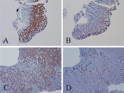
Stomach tissue samples (stained brown) to detect non-cytolytic enterovirus in patients with functional dyspepsia.
From the article:
Figure 1. Staining for VP1 and dsRNA in stomach biopsies of two functional dyspepsia patients. Panels A and B demonstrate positive staining for VP 1 with 5D8/1 mAb and dsRNA with J2 mAb, respectively for patient 1 (brown colored cells, 100× magnification); panels C and D showed the same for patient 2.
Results: At the time of EGD, careful inspection of the stomach of FD patients with or without ME/CFS, demonstrated focal or rarely diffuse, erythematous areas in the antrum in approximately 80% of patients, and less often in the body/fundus of the stomach. Microscopic examinations demonstrated minimal or mild chronic inflammation in more than 95% of biopsy samples but H. pylori was identified in less than 5% of samples by immunochemical staining.Immunoperoxidase staining demonstrated enterovirus VP1 in 343/416 (82%) and 53/66 (83%) of the stomach biopsies from FD patients with and without ME/CFS, respectively, and dsRNA in 268/ 416 (64%) and 41/65 (63%) in the two patient cohorts, respectively (Figure 1). 9/47 (19%) and 5/46 (11%) of the controls stained positive for VP1 and dsRNA, respectively (p < 0.01, χ2 test with Yates correction).
Functional dyspepsia (FD) patients with and without the diagnosis of ME/CFS, and were able to support the viral protein staining with finding of double-stranded RNA in 63% of the same stomach biopsies by immunoperoxidase staining. Furthermore, we clarified the possible cross-reaction with creatine kinase brain subtype (CKB), present in parietal cells, using antibody competition experiments and western blot analysis of stomach proteins. Viral protein+ and dsRNA+ biopsies were infectious in SCID mice. More research is needed to elucidate the mechanism of enterovirus infection of the stomach associated with FD and chronic gastritis.
Author[edit | edit source]
Chia, J.K., Chia, A. Y., Wang, D., & El-Habbal, R. (2015). Functional Dyspepsia and Chronic Gastritis Associated with Enteroviruses. Open Journal of Gastroenterology. 5(4):21.[1] Fig. 1.
doi:10.4236/ojgas.2015.54005.
Source[edit | edit source]
http://file.scirp.org/Html/2-1900264_55465.htm
 |
This file is licensed under the Creative Commons Attribution 4.0 International license. | |
|
References[edit | edit source]
- ↑ Chia, John K.; Chia, Andrew Y.; Wang, David; El-Habbal, Rabiha (April 9, 2015). "Functional Dyspepsia and Chronic Gastritis Associated with Enteroviruses". Open Journal of Gastroenterology. 05 (04): 21. doi:10.4236/ojgas.2015.54005.
File history
Click on a date/time to view the file as it appeared at that time.
| Date/Time | Thumbnail | Dimensions | User | Comment | |
|---|---|---|---|---|---|
| current | 02:20, August 8, 2018 | | 400 × 300 (273 KB) | Hip (talk | contribs) | This image it from the Chia et al 2015 study "Functional Dyspepsia and Chronic Gastritis Associated with Enteroviruses" http://file.scirp.org/Html/2-1900264_55465.htm . It shows stomach tissue samples stained (brown) to detect non-cytolytic enterovirus. |
You cannot overwrite this file.
File usage
The following 2 pages use this file: