Magnetic resonance imaging: Difference between revisions
From MEpedia, a crowd-sourced encyclopedia of ME and CFS science and history
(cat) |
(→Notable studies: add studies) |
||
| Line 20: | Line 20: | ||
== Notable studies == | == Notable studies == | ||
* 1993, A comparison of brain MRI scans from 52 CFS patients and 52 controls found that 27% of CFS patients had findings considered abnormal, while only 2% of controls had findings considered abnormal. Abnormalities included T2 hyperintensities and ventricular enlargement.<ref>{{Cite journal|last=Natelson|first=B. H.|last2=Cohen|first2=J. M.|last3=Brassloff|first3=I.|last4=Lee|first4=H. J.|date=1993-12-15|title=A controlled study of brain magnetic resonance imaging in patients with the chronic fatigue syndrome|url=https://www.ncbi.nlm.nih.gov/pubmed/8138812|journal=Journal of the Neurological Sciences|volume=120|issue=2|pages=213–217|doi=10.1016/0022-510x(93)90276-5|issn=0022-510X|pmid=8138812}}</ref> | |||
* 1999, A comparison of brain MRI scans from 39 CFS patients and 19 controls found that the 21 CFS patients who did not have a psychiatric diagnosis had significantly more T2 hyperintensities, compared to either controls or the 18 CFS patients with a psychiatric diagnosis.<ref>{{Cite journal|last=Lange|first=G.|last2=DeLuca|first2=J.|last3=Maldjian|first3=J. A.|last4=Lee|first4=H.|last5=Tiersky|first5=L. A.|last6=Natelson|first6=B. H.|date=1999-12-01|title=Brain MRI abnormalities exist in a subset of patients with chronic fatigue syndrome|url=https://www.ncbi.nlm.nih.gov/pubmed/10567042|journal=Journal of the Neurological Sciences|volume=171|issue=1|pages=3–7|doi=10.1016/s0022-510x(99)00243-9|issn=0022-510X|pmid=10567042}}</ref> | |||
*2016, Progressive Brain Changes in Patients With Chronic Fatigue Syndrome: A Longitudinal MRI Study<ref>{{Cite journal|last=Shan|first=Zack Y.|author-link=Zack Shan|last2=Kwiatek|first2=Richard|author-link2=Richard Kwiatek|last3=Burnet|first3=Richard|author-link3=Richard Burnet|last4=Fante|first4=Peter Del|author-link4=Peter Del Fante|last5=Staines|first5=Donald R.|author-link5=Donald Staines|last6=Marshall‐Gradisnik|first6=Sonya M.|author-link6=Sonya Marshall-Gradisnik|last7=Barnden|first7=Leighton R.|date=2016|title=Progressive brain changes in patients with chronic fatigue syndrome: A longitudinal MRI study|url=https://onlinelibrary.wiley.com/doi/abs/10.1002/jmri.25283|journal=Journal of Magnetic Resonance Imaging|language=en|volume=44|issue=5|pages=1301–1311|doi=10.1002/jmri.25283|issn=1522-2586|pmc=5111735|pmid=27123773|quote=|via=}}</ref> | *2016, Progressive Brain Changes in Patients With Chronic Fatigue Syndrome: A Longitudinal MRI Study<ref>{{Cite journal|last=Shan|first=Zack Y.|author-link=Zack Shan|last2=Kwiatek|first2=Richard|author-link2=Richard Kwiatek|last3=Burnet|first3=Richard|author-link3=Richard Burnet|last4=Fante|first4=Peter Del|author-link4=Peter Del Fante|last5=Staines|first5=Donald R.|author-link5=Donald Staines|last6=Marshall‐Gradisnik|first6=Sonya M.|author-link6=Sonya Marshall-Gradisnik|last7=Barnden|first7=Leighton R.|date=2016|title=Progressive brain changes in patients with chronic fatigue syndrome: A longitudinal MRI study|url=https://onlinelibrary.wiley.com/doi/abs/10.1002/jmri.25283|journal=Journal of Magnetic Resonance Imaging|language=en|volume=44|issue=5|pages=1301–1311|doi=10.1002/jmri.25283|issn=1522-2586|pmc=5111735|pmid=27123773|quote=|via=}}</ref> | ||
*2016, Autonomic correlations with MRI are abnormal in the brainstem vasomotor centre in Chronic Fatigue Syndrome<ref>{{Cite journal|last=Barnden|first=Leighton R.|author-link=Leighton Barnden|last2=Kwiatek|first2=Richard|author-link2=Richard Kwiatek|last3=Crouch|first3=Benjamin|author-link3=Benjamin Crouch|last4=Burnet|first4=Richard|author-link4=Richard Burnet|last5=Del Fante|first5=Peter|author-link5=Peter Del Fante|date=2016-01-01|title=Autonomic correlations with MRI are abnormal in the brainstem vasomotor centre in Chronic Fatigue Syndrome|url=http://www.sciencedirect.com/science/article/pii/S2213158216300584|journal=NeuroImage: Clinical|volume=11|issue=|pages=530–537|doi=10.1016/j.nicl.2016.03.017|issn=2213-1582|quote=|via=}}</ref> | *2016, Autonomic correlations with MRI are abnormal in the brainstem vasomotor centre in Chronic Fatigue Syndrome<ref>{{Cite journal|last=Barnden|first=Leighton R.|author-link=Leighton Barnden|last2=Kwiatek|first2=Richard|author-link2=Richard Kwiatek|last3=Crouch|first3=Benjamin|author-link3=Benjamin Crouch|last4=Burnet|first4=Richard|author-link4=Richard Burnet|last5=Del Fante|first5=Peter|author-link5=Peter Del Fante|date=2016-01-01|title=Autonomic correlations with MRI are abnormal in the brainstem vasomotor centre in Chronic Fatigue Syndrome|url=http://www.sciencedirect.com/science/article/pii/S2213158216300584|journal=NeuroImage: Clinical|volume=11|issue=|pages=530–537|doi=10.1016/j.nicl.2016.03.017|issn=2213-1582|quote=|via=}}</ref> | ||
| Line 25: | Line 27: | ||
*2018, Decreased Connectivity and Increased Blood Oxygenation Level Dependent Complexity in the Default Mode Network in Individuals with Chronic Fatigue Syndrome<ref>{{Cite journal|last=Shan|first=Zack Y.|author-link=Zack Shan|last2=Finegan|first2=Kevin|author-link2=Kevin Finnegan|last3=Bhuta|first3=Sandeep|author-link3=Sandeep Bhuta|last4=Ireland|first4=Timothy|author-link4=Timothy Ireland|last5=Staines|first5=Donald R.|author-link5=Donald Staines|last6=Marshall-Gradisnik|first6=Sonya M.|author-link6=Sonya Marshall-Gradisnik|last7=Barnden|first7=Leighton R.|date=Feb 2018|title=Decreased Connectivity and Increased Blood Oxygenation Level Dependent Complexity in the Default Mode Network in Individuals with Chronic Fatigue Syndrome|url=https://www.ncbi.nlm.nih.gov/pubmed/29152994|journal=Brain Connectivity|volume=8|issue=1|pages=33–39|doi=10.1089/brain.2017.0549|issn=2158-0022|pmid=29152994|quote=|via=}}</ref> | *2018, Decreased Connectivity and Increased Blood Oxygenation Level Dependent Complexity in the Default Mode Network in Individuals with Chronic Fatigue Syndrome<ref>{{Cite journal|last=Shan|first=Zack Y.|author-link=Zack Shan|last2=Finegan|first2=Kevin|author-link2=Kevin Finnegan|last3=Bhuta|first3=Sandeep|author-link3=Sandeep Bhuta|last4=Ireland|first4=Timothy|author-link4=Timothy Ireland|last5=Staines|first5=Donald R.|author-link5=Donald Staines|last6=Marshall-Gradisnik|first6=Sonya M.|author-link6=Sonya Marshall-Gradisnik|last7=Barnden|first7=Leighton R.|date=Feb 2018|title=Decreased Connectivity and Increased Blood Oxygenation Level Dependent Complexity in the Default Mode Network in Individuals with Chronic Fatigue Syndrome|url=https://www.ncbi.nlm.nih.gov/pubmed/29152994|journal=Brain Connectivity|volume=8|issue=1|pages=33–39|doi=10.1089/brain.2017.0549|issn=2158-0022|pmid=29152994|quote=|via=}}</ref> | ||
*2018, Brain function characteristics of chronic fatigue syndrome: A task fMRI study<ref>{{Cite journal|last=Shan|first=Zack Y.|author-link=Zack Shan|last2=Finegan|first2=Kevin|author-link2=Kevin Finnegan|last3=Bhuta|first3=Sandeep|author-link3=Sandeep Bhuta|last4=Ireland|first4=Timothy|author-link4=Timothy Ireland|last5=Staines|first5=Donald R.|author-link5=Donald Staines|last6=Marshall-Gradisnik|first6=Sonya M.|author-link6=Sonya Marshall-Gradisnik|last7=Barnden|first7=Leighton R.|date=2018-01-01|title=Brain function characteristics of chronic fatigue syndrome: A task fMRI study|url=http://www.sciencedirect.com/science/article/pii/S2213158218301347|journal=NeuroImage: Clinical|volume=19|issue=|pages=279–286|doi=10.1016/j.nicl.2018.04.025|issn=2213-1582|quote=|via=|author-link7=Leighton Barnden}}</ref> | *2018, Brain function characteristics of chronic fatigue syndrome: A task fMRI study<ref>{{Cite journal|last=Shan|first=Zack Y.|author-link=Zack Shan|last2=Finegan|first2=Kevin|author-link2=Kevin Finnegan|last3=Bhuta|first3=Sandeep|author-link3=Sandeep Bhuta|last4=Ireland|first4=Timothy|author-link4=Timothy Ireland|last5=Staines|first5=Donald R.|author-link5=Donald Staines|last6=Marshall-Gradisnik|first6=Sonya M.|author-link6=Sonya Marshall-Gradisnik|last7=Barnden|first7=Leighton R.|date=2018-01-01|title=Brain function characteristics of chronic fatigue syndrome: A task fMRI study|url=http://www.sciencedirect.com/science/article/pii/S2213158218301347|journal=NeuroImage: Clinical|volume=19|issue=|pages=279–286|doi=10.1016/j.nicl.2018.04.025|issn=2213-1582|quote=|via=|author-link7=Leighton Barnden}}</ref> | ||
==Learn more== | ==Learn more== | ||
*[https://www.msdmanuals.com/en-gb/professional/special-subjects/principles-of-radiologic-imaging/magnetic-resonance-imaging Magnetic Resonance Imaging] - Merck Manual | *[https://www.msdmanuals.com/en-gb/professional/special-subjects/principles-of-radiologic-imaging/magnetic-resonance-imaging Magnetic Resonance Imaging] - Merck Manual | ||
Revision as of 22:45, January 23, 2020
This article is a stub. |
Magnetic Resonance Imaging (MRI) uses magnetic fields and radio waves to produce images of thin slices of tissues. MRI scans can be used to image many different parts of the body, including the brain, joints, major organs and even the whole body.[1]
MRI scans can be used for many different purposes, e.g. to show:
- abnormalities of the brain and spinal cord
- abnormalities in various parts of the body such as breast, prostate, and liver
- joint injuries or abnormalities, for example a knee injury
- heart structure and function
- areas of activity within the brain, using a functional MRI
- blood flow through blood vessels and arteries[2]
Theory[edit | edit source]
ME/CFS MRI evidence[edit | edit source]
Cost and availability[edit | edit source]
Notable studies[edit | edit source]
- 1993, A comparison of brain MRI scans from 52 CFS patients and 52 controls found that 27% of CFS patients had findings considered abnormal, while only 2% of controls had findings considered abnormal. Abnormalities included T2 hyperintensities and ventricular enlargement.[4]
- 1999, A comparison of brain MRI scans from 39 CFS patients and 19 controls found that the 21 CFS patients who did not have a psychiatric diagnosis had significantly more T2 hyperintensities, compared to either controls or the 18 CFS patients with a psychiatric diagnosis.[5]
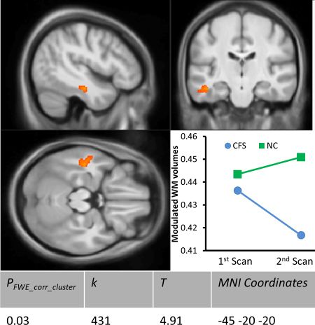
- 2016, Progressive Brain Changes in Patients With Chronic Fatigue Syndrome: A Longitudinal MRI Study[6]
- 2016, Autonomic correlations with MRI are abnormal in the brainstem vasomotor centre in Chronic Fatigue Syndrome[7]
- 2017, Medial prefrontal cortex deficits correlate with unrefreshing sleep in patients with chronic fatigue syndrome[8]
- 2018, Decreased Connectivity and Increased Blood Oxygenation Level Dependent Complexity in the Default Mode Network in Individuals with Chronic Fatigue Syndrome[9]
- 2018, Brain function characteristics of chronic fatigue syndrome: A task fMRI study[10]
Learn more[edit | edit source]
- Magnetic Resonance Imaging - Merck Manual
- MRI scans - MedlinePlus
- Head MRI - Radiology info
See also[edit | edit source]
References[edit | edit source]
- ↑ "Magnetic Resonance Imaging - Special Subjects - MSD Manual Professional Edition". MSD Manual Professional Edition. Retrieved October 12, 2018.
- ↑ Health Center for Devices and Radiological. "MRI (Magnetic Resonance Imaging) - Uses". www.fda.gov. Retrieved October 12, 2018. Cite has empty unknown parameter:
|dead-url=(help) - ↑ Shan, Zack Y.; Kwiatek, Richard; Burnet, Richard; Del Fante, Peter; Staines, Donald R.; Marshall-Gradisnik, Sonya M.; Barnden, Leighton R. (April 28, 2016). "Progressive brain changes in patients with chronic fatigue syndrome: A longitudinal MRI study". Journal of Magnetic Resonance Imaging. 44 (5): 1301–1311. doi:10.1002/jmri.25283. ISSN 1053-1807. PMC 5111735. PMID 27123773.
- ↑ Natelson, B. H.; Cohen, J. M.; Brassloff, I.; Lee, H. J. (December 15, 1993). "A controlled study of brain magnetic resonance imaging in patients with the chronic fatigue syndrome". Journal of the Neurological Sciences. 120 (2): 213–217. doi:10.1016/0022-510x(93)90276-5. ISSN 0022-510X. PMID 8138812.
- ↑ Lange, G.; DeLuca, J.; Maldjian, J. A.; Lee, H.; Tiersky, L. A.; Natelson, B. H. (December 1, 1999). "Brain MRI abnormalities exist in a subset of patients with chronic fatigue syndrome". Journal of the Neurological Sciences. 171 (1): 3–7. doi:10.1016/s0022-510x(99)00243-9. ISSN 0022-510X. PMID 10567042.
- ↑ Shan, Zack Y.; Kwiatek, Richard; Burnet, Richard; Fante, Peter Del; Staines, Donald R.; Marshall‐Gradisnik, Sonya M.; Barnden, Leighton R. (2016). "Progressive brain changes in patients with chronic fatigue syndrome: A longitudinal MRI study". Journal of Magnetic Resonance Imaging. 44 (5): 1301–1311. doi:10.1002/jmri.25283. ISSN 1522-2586. PMC 5111735. PMID 27123773.
- ↑ Barnden, Leighton R.; Kwiatek, Richard; Crouch, Benjamin; Burnet, Richard; Del Fante, Peter (January 1, 2016). "Autonomic correlations with MRI are abnormal in the brainstem vasomotor centre in Chronic Fatigue Syndrome". NeuroImage: Clinical. 11: 530–537. doi:10.1016/j.nicl.2016.03.017. ISSN 2213-1582.
- ↑ Shan, Zack Y.; Kwiatek, Richard; Burnet, Richard; Fante, Peter Del; Staines, Donald R.; Marshall‐Gradisnik, Sonya M.; Barnden, Leighton R. (2017). "Medial prefrontal cortex deficits correlate with unrefreshing sleep in patients with chronic fatigue syndrome". NMR in Biomedicine. 30 (10): e3757. doi:10.1002/nbm.3757. ISSN 1099-1492.
- ↑ Shan, Zack Y.; Finegan, Kevin; Bhuta, Sandeep; Ireland, Timothy; Staines, Donald R.; Marshall-Gradisnik, Sonya M.; Barnden, Leighton R. (February 2018). "Decreased Connectivity and Increased Blood Oxygenation Level Dependent Complexity in the Default Mode Network in Individuals with Chronic Fatigue Syndrome". Brain Connectivity. 8 (1): 33–39. doi:10.1089/brain.2017.0549. ISSN 2158-0022. PMID 29152994.
- ↑ Shan, Zack Y.; Finegan, Kevin; Bhuta, Sandeep; Ireland, Timothy; Staines, Donald R.; Marshall-Gradisnik, Sonya M.; Barnden, Leighton R. (January 1, 2018). "Brain function characteristics of chronic fatigue syndrome: A task fMRI study". NeuroImage: Clinical. 19: 279–286. doi:10.1016/j.nicl.2018.04.025. ISSN 2213-1582.


