

File:Brain-scan-CFS-basal-ganglia.png
From MEpedia, a crowd-sourced encyclopedia of ME and CFS science and history
Size of this preview: 800 × 497 pixels. Other resolution: 1,483 × 921 pixels.
Original file (1,483 × 921 pixels, file size: 714 KB, MIME type: image/png)
Summary[edit | edit source]
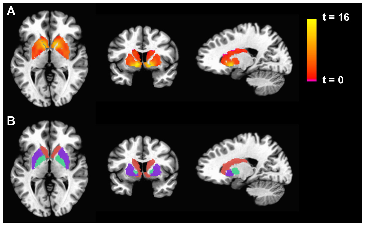
Brain scans: Chronic Fatigue Syndrome vs controls: Reduced Activation in Basal Ganglia Structures in CFS compared to Controls for the Win-Lose Contrast.
Citation:[edit | edit source]
Miller AH, Jones JF, Drake DF, Tian H, Unger ER, Pagnoni G (2014). Decreased Basal Ganglia Activation in Subjects with Chronic Fatigue Syndrome: Association with Symptoms of Fatigue. PLoS ONE 9(5): e98156. doi:10.1371/journal.pone.0098156 source: https://journals.plos.org/plosone/article/figures?id=10.1371/journal.pone.0098156
References[edit | edit source]
Licensing[edit | edit source]
 |
This file is licensed under the Creative Commons Attribution 4.0 International license. | |
|
See also[edit | edit source]
File history
Click on a date/time to view the file as it appeared at that time.
| Date/Time | Thumbnail | Dimensions | User | Comment | |
|---|---|---|---|---|---|
| current | 18:27, September 3, 2019 | | 1,483 × 921 (714 KB) | Notjusttired (talk | contribs) | Brain scans: Chronic Fatigue Syndrome vs controls: Reduced Activation in Basal Ganglia Structures in CFS compared to Controls for the Win-Lose Contrast. Citation: Miller AH, Jones JF, Drake DF, Tian H, Unger ER, Pagnoni G (2014) Decreased Basal Gangli... |
You cannot overwrite this file.
File usage
The following 2 pages use this file: