

File:Fibromyalgia-brain.jpg: Difference between revisions
Notjusttired (talk | contribs) m (Text replacement - "|first4=" to " | first4 = ") |
Notjusttired (talk | contribs) m (Text replacement - "|author-link5=" to " | authorlink5 = ") |
||
| Line 4: | Line 4: | ||
[11C]PBR28 VT was elevated in several brain regions compared to healthy control subjects (Fig. 1), including dorsolateral prefrontal cortex (dlPFC), dorsomedial PFC (dmPFC), primary somatosensory and motor cortices (S1/M1), precuneus, posterior cingulate cortex (PCC), supplementary motor area (SMA), supramarginal gyrus (SMG), and superior parietal lobule (SPL). Additionally, an ROI analysis of the aMCC revealed elevated VT in the FM patients that approached statistical significance (p = 0.071). There were no regions where control VT was significantly higher than FM VT. | [11C]PBR28 VT was elevated in several brain regions compared to healthy control subjects (Fig. 1), including dorsolateral prefrontal cortex (dlPFC), dorsomedial PFC (dmPFC), primary somatosensory and motor cortices (S1/M1), precuneus, posterior cingulate cortex (PCC), supplementary motor area (SMA), supramarginal gyrus (SMG), and superior parietal lobule (SPL). Additionally, an ROI analysis of the aMCC revealed elevated VT in the FM patients that approached statistical significance (p = 0.071). There were no regions where control VT was significantly higher than FM VT. | ||
==== Author: (or citation) ==== | ==== Author: (or citation) ==== | ||
{{Cite journal|last=Albrecht | first = Daniel S.|author-link= | last2 = Forsberg | first2 = Anton|author-link2= | last3 = Sandström | first3 = Angelica|author-link3= | last4 = Bergan | first4 = Courtney|author-link4= | last5 = Kadetoff | first5 = Diana| | {{Cite journal|last=Albrecht | first = Daniel S.|author-link= | last2 = Forsberg | first2 = Anton|author-link2= | last3 = Sandström | first3 = Angelica|author-link3= | last4 = Bergan | first4 = Courtney|author-link4= | last5 = Kadetoff | first5 = Diana | authorlink5 = |last6 = Protsenko | first6 = Ekaterina | authorlink6 = |last7 = Lampa|first7=Jon | last8 = Lee | first8 = Yvonne C.|last9 = Höglund | first9 = Caroline Olgart|date=2019-01-01|title=Brain glial activation in fibromyalgia – A multi-site positron emission tomography investigation|url=https://www.sciencedirect.com/science/article/pii/S0889159118302423|journal=Brain, Behavior, and Immunity|language=en|volume=75|issue=|pages=72–83|doi=10.1016/j.bbi.2018.09.018|issn=0889-1591|pmc=|pmid=|access-date=|quote=|via=}} | ||
Albrecht, Daniel S., et al. [https://www.sciencedirect.com/science/article/pii/S0889159118302423 "Brain glial activation in fibromyalgia–A multi-site positron emission tomography investigation."] Brain, behavior, and immunity 75 (2019): 72-83. | Albrecht, Daniel S., et al. [https://www.sciencedirect.com/science/article/pii/S0889159118302423 "Brain glial activation in fibromyalgia–A multi-site positron emission tomography investigation."] Brain, behavior, and immunity 75 (2019): 72-83. | ||
==== Source: (e.g. internet address) ==== | ==== Source: (e.g. internet address) ==== | ||
[https://www.sciencedirect.com/science/article/pii/S0889159118302423 Brain glial activation in fibromyalgia – A multi-site positron emission tomography investigation]<ref name="Albrecth2019">{{Cite journal|last=Albrecht | first = Daniel S.|author-link= | last2 = Forsberg | first2 = Anton|author-link2= | last3 = Sandström | first3 = Angelica|author-link3= | last4 = Bergan | first4 = Courtney|author-link4= | last5 = Kadetoff | first5 = Diana| | [https://www.sciencedirect.com/science/article/pii/S0889159118302423 Brain glial activation in fibromyalgia – A multi-site positron emission tomography investigation]<ref name="Albrecth2019">{{Cite journal|last=Albrecht | first = Daniel S.|author-link= | last2 = Forsberg | first2 = Anton|author-link2= | last3 = Sandström | first3 = Angelica|author-link3= | last4 = Bergan | first4 = Courtney|author-link4= | last5 = Kadetoff | first5 = Diana | authorlink5 = |last6 = Protsenko | first6 = Ekaterina | authorlink6 = |last7 = Lampa|first7=Jon | last8 = Lee | first8 = Yvonne C.|last9 = Höglund | first9 = Caroline Olgart|date=2019-01-01|title=Brain glial activation in fibromyalgia – A multi-site positron emission tomography investigation|url=https://www.sciencedirect.com/science/article/pii/S0889159118302423|journal=Brain, Behavior, and Immunity|language=en|volume=75|issue=|pages=72–83|doi=10.1016/j.bbi.2018.09.018|issn=0889-1591|pmc=|pmid=|access-date=|quote=|via=}}</ref> | ||
Fig. 1. Voxelwise group differences in [11C]PBR28 VT. A: Surface projection maps displaying areas with significantly elevated [11C]PBR28 VT in FM patients compared to controls (FM – n = 11; HC – n = 11) in voxelwise analyses (KI-only sample). B: average ± standard deviation VT extracted from several regions. The S1/M1, dLPFC and precuneus data were extracted from the clusters identified as statistically significant in the voxelwise VT analysis. For these regions, the plots are displayed for illustrative purposes only, and the level of statistical significance noted for each plot reflects that of the voxelwise analyses. For the aMCC, the data was extracted from a region independently identified based on the results of the SUVR voxelwise analysis (see Fig. 2). | Fig. 1. Voxelwise group differences in [11C]PBR28 VT. A: Surface projection maps displaying areas with significantly elevated [11C]PBR28 VT in FM patients compared to controls (FM – n = 11; HC – n = 11) in voxelwise analyses (KI-only sample). B: average ± standard deviation VT extracted from several regions. The S1/M1, dLPFC and precuneus data were extracted from the clusters identified as statistically significant in the voxelwise VT analysis. For these regions, the plots are displayed for illustrative purposes only, and the level of statistical significance noted for each plot reflects that of the voxelwise analyses. For the aMCC, the data was extracted from a region independently identified based on the results of the SUVR voxelwise analysis (see Fig. 2). | ||
Revision as of 01:23, December 1, 2022
Summary[edit | edit source]
Title: (or description)[edit | edit source]
Brain abnormalities in fibromyalgia.
[11C]PBR28 VT was elevated in several brain regions compared to healthy control subjects (Fig. 1), including dorsolateral prefrontal cortex (dlPFC), dorsomedial PFC (dmPFC), primary somatosensory and motor cortices (S1/M1), precuneus, posterior cingulate cortex (PCC), supplementary motor area (SMA), supramarginal gyrus (SMG), and superior parietal lobule (SPL). Additionally, an ROI analysis of the aMCC revealed elevated VT in the FM patients that approached statistical significance (p = 0.071). There were no regions where control VT was significantly higher than FM VT.
Author: (or citation)[edit | edit source]
Albrecht, Daniel S.; Forsberg, Anton; Sandström, Angelica; Bergan, Courtney; Kadetoff, Diana; Protsenko, Ekaterina; Lampa, Jon; Lee, Yvonne C.; Höglund, Caroline Olgart (January 1, 2019). "Brain glial activation in fibromyalgia – A multi-site positron emission tomography investigation". Brain, Behavior, and Immunity. 75: 72–83. doi:10.1016/j.bbi.2018.09.018. ISSN 0889-1591. Albrecht, Daniel S., et al. "Brain glial activation in fibromyalgia–A multi-site positron emission tomography investigation." Brain, behavior, and immunity 75 (2019): 72-83.
Source: (e.g. internet address)[edit | edit source]
Brain glial activation in fibromyalgia – A multi-site positron emission tomography investigation[1]
Fig. 1. Voxelwise group differences in [11C]PBR28 VT. A: Surface projection maps displaying areas with significantly elevated [11C]PBR28 VT in FM patients compared to controls (FM – n = 11; HC – n = 11) in voxelwise analyses (KI-only sample). B: average ± standard deviation VT extracted from several regions. The S1/M1, dLPFC and precuneus data were extracted from the clusters identified as statistically significant in the voxelwise VT analysis. For these regions, the plots are displayed for illustrative purposes only, and the level of statistical significance noted for each plot reflects that of the voxelwise analyses. For the aMCC, the data was extracted from a region independently identified based on the results of the SUVR voxelwise analysis (see Fig. 2).
Other information:[edit | edit source]
Licensing[edit | edit source]

|
|
This work is licensed under the Creative Commons Attribution-NonCommercial No-Derivatives 4.0 International license.
|


- ↑ Albrecht, Daniel S.; Forsberg, Anton; Sandström, Angelica; Bergan, Courtney; Kadetoff, Diana; Protsenko, Ekaterina; Lampa, Jon; Lee, Yvonne C.; Höglund, Caroline Olgart (January 1, 2019). "Brain glial activation in fibromyalgia – A multi-site positron emission tomography investigation". Brain, Behavior, and Immunity. 75: 72–83. doi:10.1016/j.bbi.2018.09.018. ISSN 0889-1591.
File history
Click on a date/time to view the file as it appeared at that time.
| Date/Time | Thumbnail | Dimensions | User | Comment | |
|---|---|---|---|---|---|
| current | 20:50, November 27, 2021 | 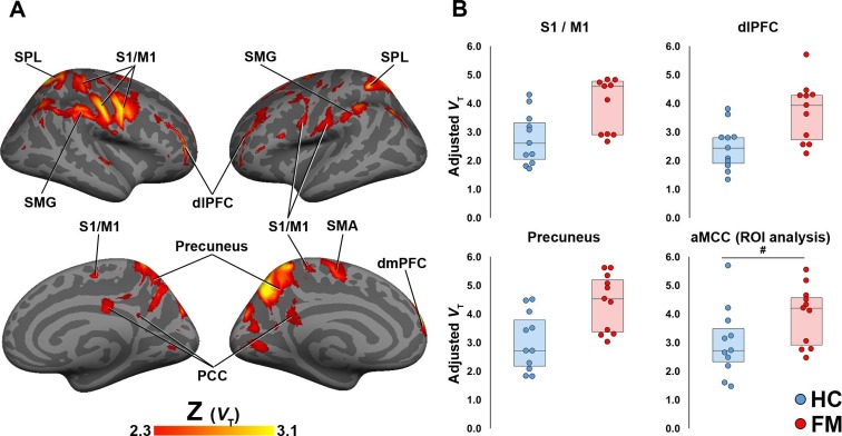 | 756 × 392 (77 KB) | Notjusttired (talk | contribs) | ==== Title: (or description) ==== Brain abnormalities in fibromyalgia. In FM patients, [11C]PBR28 VT was elevated in several brain regions compared to healthy control subjects (Fig. 1), including dorsolateral prefrontal cortex (dlPFC), dorsomedial... |
You cannot overwrite this file.
File usage
There are no pages that use this file.