

File:Dorsal Root Ganglion.JPG: Difference between revisions
Notjusttired (talk | contribs) m (Text replacement - "|first4=" to " | first4 = ") |
Notjusttired (talk | contribs) m (Text replacement - "|last3=" to " | last3 = ") |
||
| Line 11: | Line 11: | ||
Source: [http://virtualslides.med.umich.edu/Histology/Basic%20Tissues/Nervous%20Tissue/065-2_HISTO_40X.svs/view.apml the Regents of University of Michigan Medical School ©2012], cropped and labelled in {{Cite book| | Source: [http://virtualslides.med.umich.edu/Histology/Basic%20Tissues/Nervous%20Tissue/065-2_HISTO_40X.svs/view.apml the Regents of University of Michigan Medical School ©2012], cropped and labelled in {{Cite book| | ||
last=Betts | first=JG|authorlink= | last2 = Young | first2 = KA | authorlink2 = |last3=Wise | first3 = JA | last4 = Johnson | first4 = E | last5 = Poe | first5 = B|last6 = Kruse | first6 = DH | last7 = Korol | first7 = O|last8 = Johnson | first8 = JE|last9 = Womble|first9 = M | last10 = DeSaix | first10 = P|date=Apr 25, 2013 | last=Betts | first=JG|authorlink= | last2 = Young | first2 = KA | authorlink2 = | last3 = Wise | first3 = JA | last4 = Johnson | first4 = E | last5 = Poe | first5 = B|last6 = Kruse | first6 = DH | last7 = Korol | first7 = O|last8 = Johnson | first8 = JE|last9 = Womble|first9 = M | last10 = DeSaix | first10 = P|date=Apr 25, 2013 | ||
|website=OpenStax|location=Houston, Texas|language=en|archive-url=|archive-date=|url-status= | |website=OpenStax|location=Houston, Texas|language=en|archive-url=|archive-date=|url-status= | ||
|chapter=13.4 The Peripheral Nervous System | title =Anatomy and Physiology | url=https://openstax.org/books/anatomy-and-physiology/pages/1-introduction | |chapter=13.4 The Peripheral Nervous System | title =Anatomy and Physiology | url=https://openstax.org/books/anatomy-and-physiology/pages/1-introduction | ||
Revision as of 01:22, December 1, 2022
Title:[edit | edit source]
Dorsal root ganglion. The cell bodies of sensory neurons, which are unipolar neurons by shape, are seen in this photomicrograph. Also, the fibrous region is composed of the axons of these neurons that are passing through the ganglion to be part of the dorsal nerve root (tissue source: canine). LM × 40.
Author:[edit | edit source]
The Regents of University of Michigan Medical School, with labelling and cropping by OpenStax
Source:[edit | edit source]
©The Regents of University of Michigan Medical School; Michigan Histology and Virtual Microscopy Learning Resources (2012). "Micrograph of dorsal root ganglion". Retrieved January 27, 2022 – via Betts, JG; Young, KA; Wise, JA; Johnson, E; Poe, B; Kruse, DH; Korol, O; Johnson, JE; Womble, M; DeSaix, P (April 25, 2013). "13.4 The Peripheral Nervous System". Anatomy and Physiology. OpenStax. Houston, Texas.
Text for use in wiki pages in the image caption[edit | edit source]
Source: the Regents of University of Michigan Medical School ©2012, cropped and labelled in Betts, JG; Young, KA; Wise, JA; Johnson, E; Poe, B; Kruse, DH; Korol, O; Johnson, JE; Womble, M; DeSaix, P (April 25, 2013). "13.4 The Peripheral Nervous System". Anatomy and Physiology. OpenStax. Houston, Texas. Retrieved January 27, 2022.. License: CC BY-NC-SA 4.0.
License:[edit | edit source]
CC BY-NC-SA 4.0, U Michigan confirmation. Template:CC-by-nc-sa 4.0
| Used with permission: This file has been uploaded with the creator's permission. It may not be licensed for others to reuse, or the copyright owner may have given unusual permission, e.g. non-commercial permission without a specific license, or permission for MEpedia only. See MEpedia:Copyright policy for more information. |
More information[edit | edit source]
View the University of Michigan WebScope at http://virtualslides.med.umich.edu/Histology/Basic%20Tissues/Nervous%20Tissue/065-2_HISTO_40X.svs/view.apml to explore the tissue sample in greater detail.
File history
Click on a date/time to view the file as it appeared at that time.
| Date/Time | Thumbnail | Dimensions | User | Comment | |
|---|---|---|---|---|---|
| current | 23:05, July 19, 2018 | 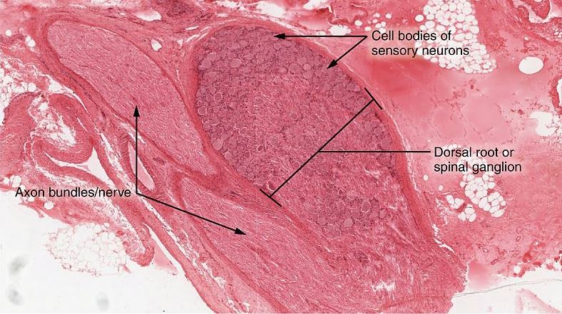 | 788 × 441 (109 KB) | MEcfsFMS (talk | contribs) | Dorsal Root Ganglion Source: Wiki Commons |
You cannot overwrite this file.
File usage
The following page uses this file: